One of the major arteries in the human body is Femoral Artery. It helps in the supply of blood to the lower limb. Near the groin region, this artery forms a delta shape when it passes through the femoral vein and femoral nerve. For surgeons, the femoral triangle or Scarpa’s triangle provides as a crucial anatomical landmark when surgery is require in the region. In this blog, we are going to explain all about femoral artery, deep femoral artery, and other concepts related to them. Let’s get started:
This artery is used by embalmers to deliver chemicals to the body to maintain it after death. The femoral artery is sub-divided into a superficial artery, common artery, and deep artery. All of these arteries provide blood to the different sections of the body. Profunda femoris is the largest branch of the femoral artery that supplies blood to the thigh and buttock area. To bring oxygen-depleted blood from these areas back to the heart, the femoral vein runs with this artery.
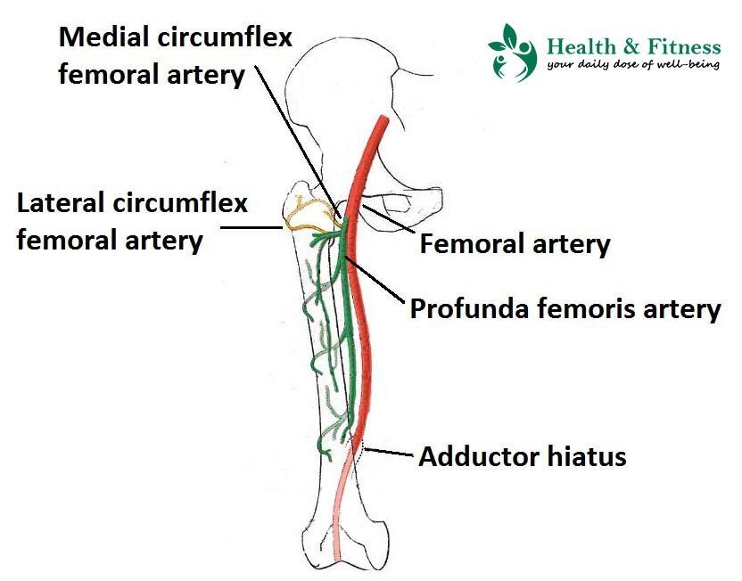
The common femoral artery continues as the superficial femoral artery and gives off the deep femoral branch.
Within the femoral triangle
In clinical practice, the relationship of the femoral artery to other structures inside the thigh can be essential. Inside the femoral triangle, the femoral artery is situated deep to the:
- Superficial fascia
- Skin
- Fascia lata
- Superficial inguinal lymph nodes
- The genitofemoral nerve’s femoral branch
- Superficial circumflex iliac vein
The medial femoral cutaneous nerve passes the artery in a lateral towards a medial direction, at the apex of the femoral triangle. Inside the triangle, the tendons of the psoas major, adductor Longus and pectineus cross deep to the femoral artery. Adjacently, at the apex of the triangle, the vein is identified deep to the artery.
To remember the content order of the femoral triangle, you can use mnemonic NAVY, from the lateral to medial:
- Nerve
- Artery
- Vein
- lYmphatics (femoral canal)
Within the adductor canal
Inside the adductor canal, the femoral artery is found deep to the:
- Superficial fascia
- Skin
- Deep fascia
- Sartorius muscle
The artery is superficial to the Longus and adductor Magnus muscles. Both nerves and veins change in their location concerning the femoral artery. The saphenous nerve or vein is found lateral to the femoral artery, however, it is also found anterior and then medial to the vein as it passes through the canal. The vastus medialis muscle and its vein are found anterolateral to the femoral artery.
Deep Femoral Artery
The branch of the femoral artery is the deep femoral artery of the human body. The common femoral artery is the largest artery in the human body that possesses multiple branches. The deep femoral artery supplies blood to the skin of the medial thigh region and muscles that flex, extend, and adduct the thigh.
It possesses oxygen-rich blood to the muscles of the thigh and upper leg, a vein eliminate oxygen-depleted blood from the thigh. From the common femoral artery, the deep femoral artery is branching off at a point referred to as the femoral triangle. Once the deep femoral artery left the femoral triangle, it starts developing further branches to deliver blood to the back of the thigh.
The medial and lateral circumflex femoral arteries, both these branches, and the deep femoral artery are essential suppliers of blood to the thigh and bones joined with it. Despite this, the medial circumflex supplies the femur with blood.
The deep femoral artery releases several other branches:
It is the first limb or branch of the deep femoral artery. It terminates the anterior element/aspect of the proximal femur by categorizing it into three branches: ascending, traverse, and descending branches. These all branches give supply for the proximal aspect of the femur, the adjacent portion of the skin of the thigh, and the quadriceps femoris muscle.
It moves around the posterior aspect of the femur where it is divided into two terminal branches: ascending and traverse. Basically, these branches or limbs give supply to the adductors of the thigh.
Perforating Femoral Arteries
These arteries perforate the proximal part of the adductor Magnus muscle so it can appear in the flexor section of the thigh. The upper three pierce or perforators are referred to as the true collateral branches of the deep femoral artery. But the fourth pierce is referred to as the terminal branch of the deep femoral artery.
Conclusion
After reading this blog, you’ll get detailed information about the femoral artery and other relate aspects. Now, you have an idea, how the femoral artery works and what exactly it is.
The femoral artery is important because it is a common site of peripheral arterial disease (PAD) complications. And, it can lead to intermittent claudication symptoms in your thigh.